
Active Perforin 1 (PRF1)
FLH2; HPLH2; P1; PFP; Pore Forming Protein; Cytolysin; Lymphocyte pore-forming protein
- Product No.APB317Hu01
- Organism SpeciesHomo sapiens (Human) Same name, Different species.
- Buffer Formulation20mM Tris, 150mM NaCl, pH8.0, containing 1mM EDTA, 1mM DTT, 0.01% SKL, 5% Trehalose and Proclin300.
- TraitsFreeze-dried powder
- Purity> 95%
- Isoelectric Point7.7
- ApplicationsCell culture; Activity Assays.
- Download Instruction Manual
- UOM 10µg50µg 200µg 1mg 5mg
-
FOB
US$ 300
For more details, please contact local distributors!US$ 750
For more details, please contact local distributors! US$ 1500
For more details, please contact local distributors! US$ 4500
For more details, please contact local distributors! US$ 11250
For more details, please contact local distributors!
-
 Packages (Simulation)
-
Packages (Simulation)
-
Figure . Gene Sequencing (extract)
-
Figure. SDS-PAGE
-
Figure. Western Blot
-
ISO9001: 2008, ISO13485: 2003 Registered
ACTIVITY TEST of the Active Perforin 1 (PRF1)

Figure. The binding activity of PRF1 with CRT.
Perforin 1 (PRF1) is a pore forming cytolytic protein found in the granules of cytotoxic T lymphocytes (CTLs) and NK cells. Upon degranulation, perforin binds to the target cell's plasma membrane, and oligomerises in a Ca2 dependent manner to form pores on the target cell. The pore formed allows for the passive diffusion of a family of pro-apoptotic proteases, known as the granzymes, into the target cell. Besides, Calreticulin (CRT) has been identified as an interactor of PRF1, thus a binding ELISA assay was conducted to detect the interaction of recombinant human PRF1 and recombinant human CRT. Briefly, PRF1 were diluted serially in PBS, with 0.01% BSA (pH 7.4). Duplicate samples of 100μL were then transferred to CRT-coated microtiter wells and incubated for 2h at 37℃. Wells were washed with PBST and incubated for 1h with anti-PRF1 pAb, then aspirated and washed 3 times. After incubation with HRP labelled secondary antibody, wells were aspirated and washed 3 times. With the addition of substrate solution, wells were incubated 15-25 minutes at 37℃. Finally, add 50µL stop solution to the wells and read at 450nm immediately. The binding activity of PRF1 and CRT was shown in Figure 1, and this effect was in a dose dependent manner.
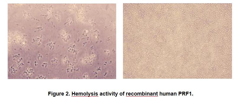
Figure. Hemolysis activity of recombinant human PRF1.
The activity of recombinant PRF1 was measured by lysis of erythrocytes using a hemolysis assay. A general procedure is as fllows: two-fold dilute the recombinant human PRF1 with 0.9% NaCl, add 50μL a serial dilution of PRF1, 10μL 0.1M CaCl2 to each well, then add 50μL 0.25% rabbit erythrocyte (RaE) to each well and mixed gently. Add 10μL 0.9% NaCl to reaplace CaCl2 in control wells. The plate is incubated for 20 hours at 37℃, 5% CO2. The results are shown in Figure 2. It was obvious that the minimal effective concentration of PRF1 is 12.5μg/mL.
(A) 0.25% RaE tread with 12.5μg/mL PRF1 for 20h;
(B) Negative control (0.25% RaE tread with 12.5ug/mL PRF1) without CaCl2.
USAGE of the Active Perforin 1 (PRF1)
Reconstitute in 20mM Tris, 150mM NaCl (pH8.0) to a concentration of 0.1-1.0 mg/mL. Do not vortex.
STORAGE of the Active Perforin 1 (PRF1)
Avoid repeated freeze/thaw cycles. Store at 2-8°C for one month. Aliquot and store at -80°C for 12 months.
STABILITY of the Active Perforin 1 (PRF1)
The thermal stability is described by the loss rate. The loss rate was determined by accelerated thermal degradation test, that is, incubate the protein at 37°C for 48h, and no obvious degradation and precipitation were observed. The loss rate is less than 5% within the expiration date under appropriate storage condition.
INCREMENT SERVICES
BCA Protein Quantification Kit
Molecular Mass Marker for Protein
Monoclonal Antibody Customized Service
Polyclonal Antibody Customized Service
Protein Activity Test Experiment Service
Electrophoretic Mobility Shift Assay (EMSA) Experiment Service
Buffer
Lentivirus Packaging Experiment Service
Adenovirus Packaging Experiment Service
Real Time PCR Experimental Service
Spike RBD Protein (S-RBD)
Protein G
Protein A
Related products
| Catalog No. | Organism species: Homo sapiens (Human) | Applications (RESEARCH USE ONLY!) |
| RPB317Hu02 | Recombinant Perforin 1 (PRF1) | Positive Control; Immunogen; SDS-PAGE; WB. |
| APB317Hu01 | Active Perforin 1 (PRF1) | Cell culture; Activity Assays. |
| RPB317Hu01 | Recombinant Perforin 1 (PRF1) | Positive Control; Immunogen; SDS-PAGE; WB. |
| APB317Hu02 | Active Perforin 1 (PRF1) | Cell culture; Activity Assays. |
| PAB317Hu01 | Polyclonal Antibody to Perforin 1 (PRF1) | WB; IHC; ICC; IP. |
| LAB317Hu81 | FITC-Linked Polyclonal Antibody to Perforin 1 (PRF1) | WB; IHC; ICC; IF. |
| LAB317Hu71 | Biotin-Linked Polyclonal Antibody to Perforin 1 (PRF1) | WB; IHC; ICC. |
| MAB317Hu22 | Monoclonal Antibody to Perforin 1 (PRF1) | WB; IHC; ICC; IP. |
| MAB317Hu21 | Monoclonal Antibody to Perforin 1 (PRF1) | WB; IHC; ICC; IP. |
| SEB317Hu | ELISA Kit for Perforin 1 (PRF1) | Enzyme-linked immunosorbent assay for Antigen Detection. |
| LMB317Hu | Multiplex Assay Kit for Perforin 1 (PRF1) ,etc. by FLIA (Flow Luminescence Immunoassay) | FLIA Kit for Antigen Detection. |